


Echocardiography, often called an echo, is a non-invasive imaging technique that uses sound waves to create detailed pictures of your heart. It allows us to visualise the heart’s chambers, valves, and surrounding structures, providing valuable information about its function. This service is crucial for individuals experiencing chest pain, shortness of breath, palpitations, or those with known heart conditions such as heart valve disease, heart failure, or congenital heart defects. It’s important because it helps us assess the heart’s pumping ability, identify structural abnormalities, and guide treatment decisions.
Procedure Overview
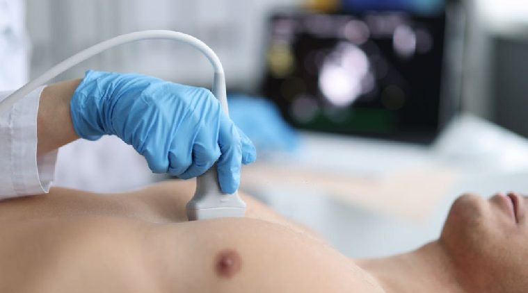
During an echocardiogram, you’ll lie on your left side or back on an examination table. A gel will be applied to your chest to help the ultrasound transducer glide smoothly over the skin. The transducer emits high-frequency sound waves that bounce off the heart, creating real-time images that are displayed on a monitor. The technician will move the transducer to capture different views of the heart. The procedure typically takes about 30-60 minutes. We utilise advanced echocardiography equipment, including Doppler technology, which allows us to assess blood flow within the heart, and 3D echocardiography, which provides detailed anatomical information.
Benefits & Importance
Echocardiography offers several advantages over other imaging methods. It’s non-invasive, meaning there’s no need for needles or radiation exposure. It provides real-time images of the heart in motion, allowing for immediate assessment of its function. It’s a valuable tool for diagnosing a wide range of heart conditions, from valve problems to heart failure. By providing detailed information about the heart’s structure and function, it helps guide treatment decisions and improve long-term heart health.
Preparation & Requirements
No specific preparation is needed for an echocardiogram. You can eat, drink, and take your medications as usual. It’s important to wear comfortable clothing that allows easy access to your chest. During the procedure, you’ll be asked to remain still while the technician captures images of your heart. After the echocardiogram, you can resume your normal activities immediately.
Risks & Considerations
Echocardiography is generally safe and well-tolerated. There are no known risks or side effects associated with the procedure. It’s a painless and non-invasive test. In rare cases, some people might experience mild discomfort from the pressure of the transducer on the chest. There are no contraindications or limitations to the procedure.
Understanding the structure and function of your heart is crucial for maintaining good cardiovascular health. An echocardiogram provides a non-invasive and accurate way to assess your heart and guide treatment decisions. If you’re experiencing chest pain, shortness of breath, palpitations, or have a known heart condition, don’t hesitate.